
Figure 1. Cartoon of a female femoral bone.
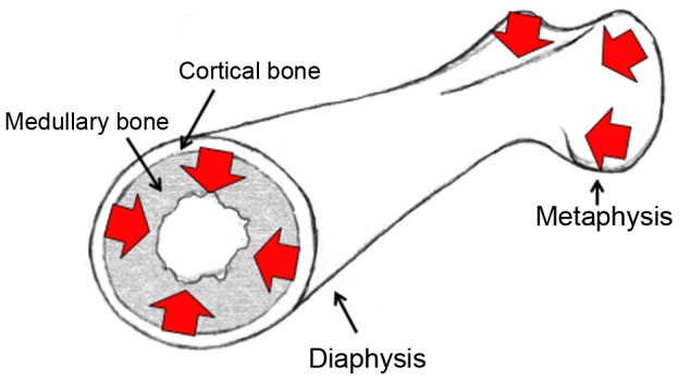
The location of medullary and cortical bone is highlighted, as well as the metaphysis and diaphysis. The flow of calcium in egg-laying females is indicated with red arrows, illustrating the transfer of calcium from the metaphyses to the diaphysis, and from the hard cortical bone to the soft medullary bone.