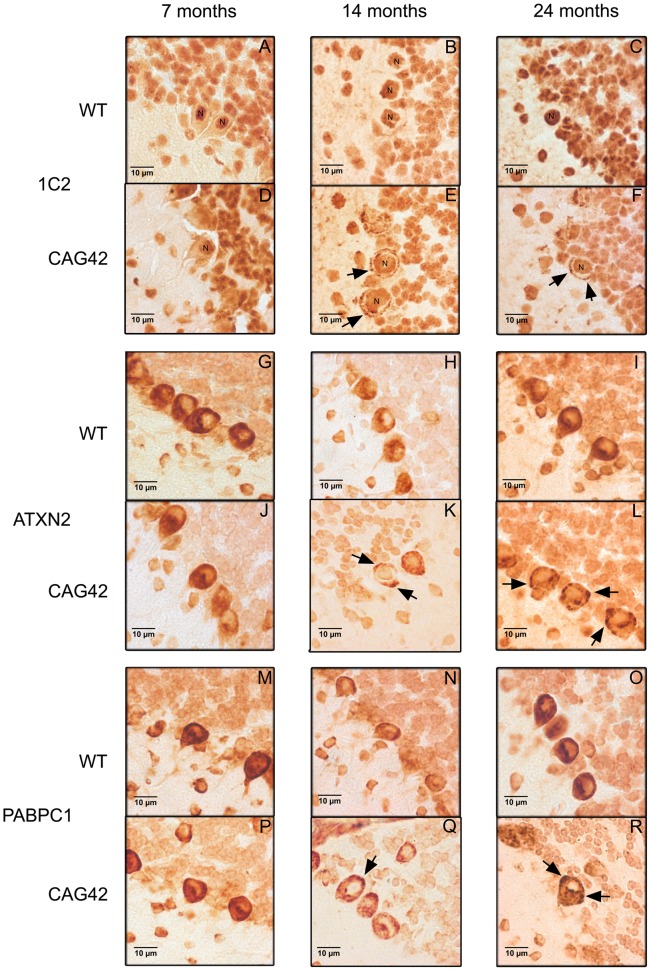
Figure 6. Immunohistochemistry of cerebellar Purkinje cells.
At 7, 14 and 24 months age, the Purkinje cells showed a purely nuclear distribution of large polyQ-domains as revealed by 1C2-staining in WT tissue, while an additional granular cytoplasmic staining pattern became apparent at 14 and 24 months in knock-in tissue (A–F). ATXN2 immunoreactivity was diffusely distributed throughout the cytoplasm and concentrated in the perinuclear region in WT tissue, while a more granular appearance was detected discretely at 14 and markedly at 24 months in CAG42 mice (G–L). The expected diffuse cytoplasmic distribution of PABPC1 was documented in WT mice; whereas again a more granular staining pattern in CAG42 mice appeared by 14 months (M–R).

