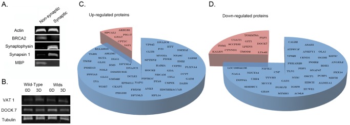
Figure 1. Proteomic identification and refinement of molecular pathways underlying degeneration in synapse-enriched brain fractions.
A. Representative bands from fluorescent western blots demonstrating enrichment of two distinct synaptic proteins (synaptophysin and synapsin-1) and relative purity of the synapse-enriched fractions, comparatively free from nuclear contamination (BRCA2) and glial cell contamination (MBP) relative to non-synaptic fractions. Actin is shown as a loading control. B. Representative bands from fluorescent western blots showing examples of protein expression changes (VAT1 and DOCK7) present in striatal synapse-enriched fractions from both wild-type and WldS mice before (0D) and 3 days after (3D) cortical lesion. Both proteins showed similar alterations in expression in degenerating tissue across wild-type and WldS mice suggesting that they are more likely to represent systemic responses to injury rather than direct mediators of the degenerative process (as synaptic degeneration is yet to be initiated in WldS mice 3 days after lesion [14]). Tubulin is shown as a loading control. C/D. Pie charts representing all proteins found to have altered expression >20% following cortical lesion. The blue section of each chart represents those proteins altered only in wild-type mice. The red sections show those proteins found to be altered in both wild-type and WldS mice, which were subsequently subtracted from the proteomic profile as they were not considered to represent expression changes underlying degeneration.

