
Figure 5. Overview of putative axo-synaptic degeneration phenotypes observed in Drosophila neurodegeneration screens.
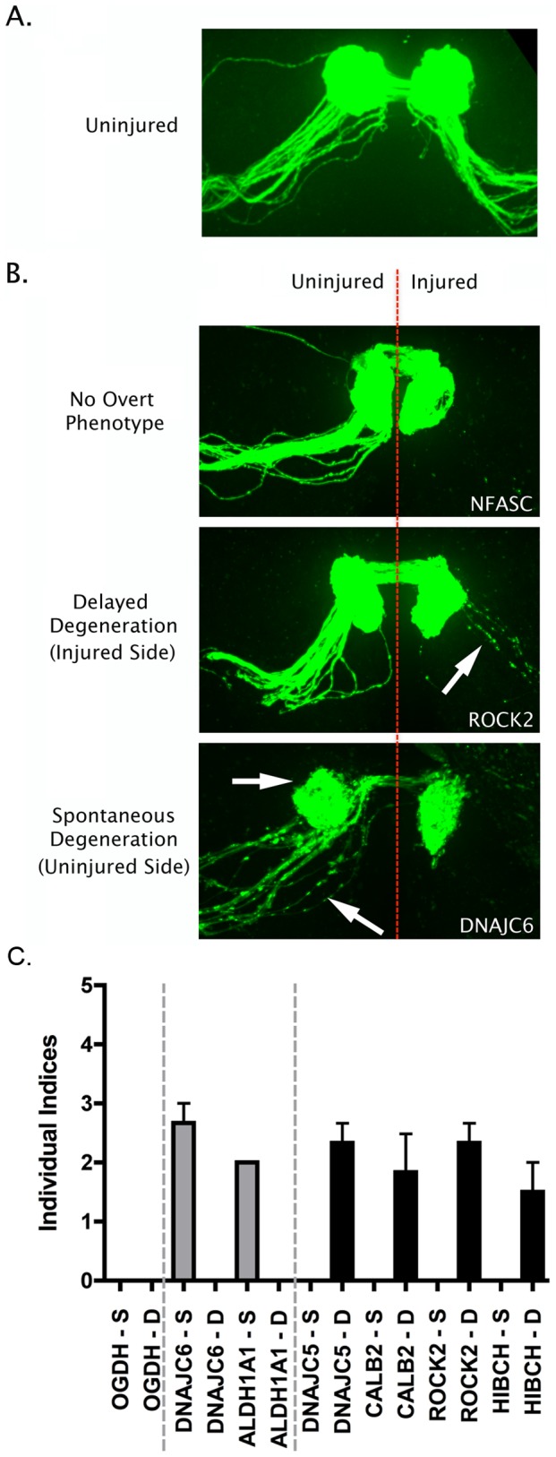
A. Representative confocal micrograph showing the morphology of the intact Drosophila olfactory receptor neuron (ORN) system, with axons and synaptic fields labeled with GFP in the UAS-mCD8::GFP,OR22a-Gal4/+ background. Axons enter the antennal lobe laterally and project medially across the lobe to reach their target glomerulus, where synapses are located (see reference [21]). B. Representative confocal micrographs showing three distinct phenotypic profiles observed in injured and un-injured ORN axons and synapses 7 days after unilateral (right hand side of image) antennal ablation. The top panel shows intact healthy axons and synapses on the uninjured side and complete axonal degeneration (indicated by absence of GFP labeled profiles) on the injured side (example from an NFASC mutant). The middle panel shows delayed axo-synaptic degeneration on the injured side, as indicated by the retention of GFP-labelled axon profiles 7 days after injury (white arrow; example from a ROCK2 mutant). The bottom panel shows spontaneous (i.e. not injury-induced) axo-synaptic degeneration in the uninjured axons and synapses, indicated by reduction and fragmentation of GFP labeled axons and synapses (white arrows; example from a DNAJC6 mutant). C. Bar chart (mean±SEM) showing index scores (see methods) for spontaneous degeneration (S; grey bars) and delayed degeneration (D; black bars) in 7 mutant Drosophila lines. OGDH is shown as an example of a mutant line with no overt phenotype. DNAJC6 and ALDH1A1 mutants revealed evidence for spontaneous degeneration in the absence of any injury stimulus. DNAJC5, CALB2, ROCK2 and HIBCH mutants revealed evidence for delayed degeneration following antennal ablation.