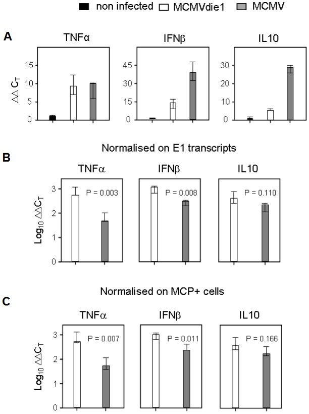
Figure 7. In situ activation of cellular genes TNFα, IFNβ and IL10 during infection of the liver.
Immune compromised female BALB/c mice (n = 3 per group) were infected with 1×10⁵ PFU of MCMV or MCMVdie1 or not infected, i.e. uninfected but immune compromised mice to take into account that a 6.5 Gy total-body γ-irradiation by itself slightly stimulates the expression of the genes under investigation. The analysis was performed 10 days after infection. (A) Expression levels (ΔΔCT values) relative to β-actin transcripts. (B, C) Normalisation of the relative expression levels of TNFα, IFNβ, and IL10 to the numbers of E1 transcripts per 500 ng of total RNA (B) or to the numbers of infected MCP+ cells per representative 10-mm2 areas of liver tissue sections (C) in order to take account of differences in tissue infection density. Throughout, bars represent median values for three mice per experimental group. Variance bars indicate the range. P values for significance are indicated for group comparisons of interest (unpaired t-test, two-sided, performed with log-transformed data).

