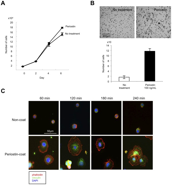
Figure 4. Periostin promotes proliferation and migration of lymphatic endothelial cells.
(A) Proliferation of TR-LE cells after recombinant periostin treatment. Cells were seeded onto fibronectin-coated 24-well plates at 1.5×10⁴/well. After pre-incubation at 33°C for 24 h, the temperature was changed and recombinant periostin (100 ng/mL) was added to the medium. The cells were trypsinized and counted 0, 2, 4, or 6 days after the addition of recombinant periostin. The bars show the average values and SDs from 3 independent experiments. (B) The effect of periostin on cell migration of TR-LE cells. Cells were seeded onto filters pre-coated with 10 µg of fibronectin. The lower compartment contained 0.5 mL of serum-free medium with or without 100 ng/mL recombinant periostin. After incubation for 4 h, cells that had migrated to the lower surfaces of the filters were visualized by hematoxylin staining and counted. The assay was repeated 3 times. The figure shows cells that had migrated to the lower surface of the filter (upper panel). The graph shows the number of cells on the lower surfaces of the filters with or without periostin (100 ng/mL) (lower panel). (C) TR-LE cells were seeded on cover slips coated with PBS or recombinant periostin (200 ng/mL) and allowed to attach for 60, 120, 180, or 240 min. The cells were stained with Alexa Fluor 488-phalloidin antibody and anti-vinculin-FITC antibody. DNA was visualized by 4′,6-diamidino-2-phenylindole (DAPI) staining.

