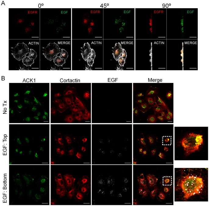
Figure 4. Cortactin localizes with ACK1 in vesicles containing ligand-bound EGFR.
(A) 1483 cells serum starved for 16 h were stimulated with 100 nanograms/milliter Alexa Fluor-488 conjugated EGF (green) for 30 min. Cells were fixed and labeled with phalloidin (Actin; pseudocolored white) and anti-EGFR antibodies (pseudocolored red). Cells were evaluated by confocal microscopy and images rotated 45° and 90° as indicated to demonstrate EGF/EGFR colocalization throughout the z-plane. (B) Serum starved (No Tx) 1483 cells were stimulated with FITC-EGF (pseudocolored white) as in (A). Cells were fixed and labeled with anti-ACK1 (green) and cortactin (red) antibodies. Confocal images of labeled EGR in the apical (top) and ventral (bottom) cellular regions are shown. Dashed boxes in the merged images indicate the areas enlarged in the photos to the right. Scale bars, 20 micrometers.

