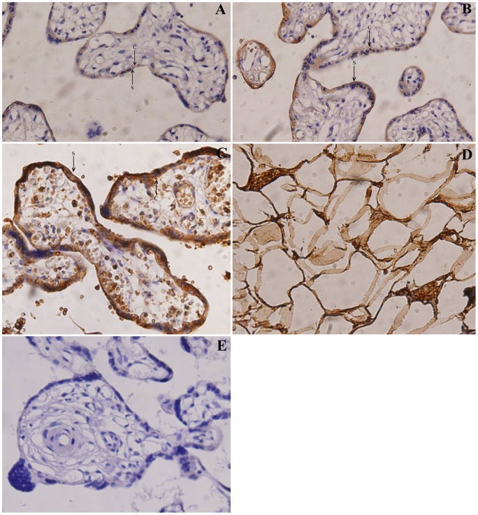
Figure 1. Immunoreactivity for OPG on human placental tissues.
Immunoreactivity for OPG on normal term placentas (A), mild preeclampsia placentas (B), and severe preeclampsia placentas (C). OPG was localized in cytotrophoblast [C] and syncytiotrophoblast [S] cells (×400). Positive control of adipose tissues shows OPG immunostaining (D; ×400); however, negative control of the primary antibody normal term placenta obtained by substituting the primary antibody with phosphate buffer saline (PBS) shows no OPG immunostaining (E; ×400).

