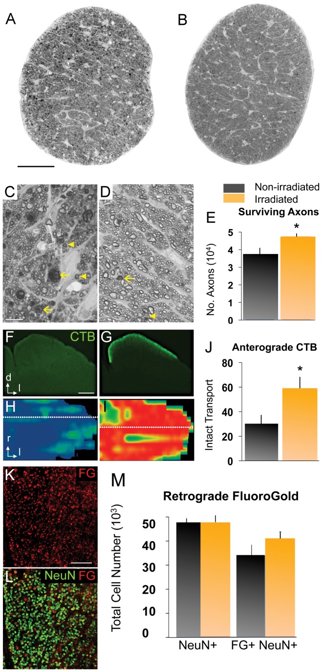
Figure 4. Head irradiation protects D2 mice from optic nerve degeneration.
(A, B) Optic nerve cross-sections stained with PPD, from naïve (11 mo old) and irradiated (12 mo old) D2 mice. Scale bar, 250 µm. (C, D) Higher magnification view of the same nerves. Scale bar, 10 µm. (C) Non-irradiated D2 nerves show abundant degenerating axon profiles with dark, condensed axoplasm (arrowheads) and disorganized myelin sheaths (arrows), as well as extensive gliosis. (D) Irradiated D2 nerves mostly show healthy axons with clear axoplasm and uniform myelin sheaths, and rare degenerating profiles (arrows). (E) Total optic axon counts from irradiated D2 nerves (n = 29; 12 mo old) show a significant increase in mean axon number compared to non-irradiated D2 mice (n = 16; 10 and 11 mo old; p<0.01). (F–J) The anterograde transport of CTB by RGC axons from the retina to the superior colliculus (SC) was measured in non-irradiated (n = 16; 10 and 11 mo old) and irradiated D2 mice (n = 17; 12 mo old). (F) Non-irradiated D2 mice (11 mo old) showed severe loss of CTB signal, as seen in coronal section of the SC (dorsal, d; lateral, l). Scale bar, 500 µm. (G) Irradiated D2 mice (12 mo old) showed persistent CTB transport to the superficial layers of the SC. (H, I) Reconstruction of the retinotopic SC map, showing the density of CTB signal (red, green and blue indicate 100, 50 and 0% density, respectively). Dotted lines indicate location of sections shown in F and G; rostral, r; lateral, l. (H) Non-irradiated D2 mice showed near total CTB depletion. (I) Irradiated D2 mice showed almost complete CTB label and a normal retinotopic pattern. (J) Irradiation significantly protected axonal anterograde transport in D2 optic nerves (p<0.05). K) Flat-mount of an irradiated D2 retina at 9 mo of age showing FG labeled cells (red). L) Most NeuN+ RGCs (green) in this retina are retrogradely labeled with FG (red). Scale bar, 100 µm. M) Quantitative stereology of both NeuN+ RGCs and FG+/NeuN+ RGCs in flat-mounted retinas. Non-irradiated retinas (dark bars, n = 8; 9–10 mo old) and irradiated retinas (yellow bars, n = 8; 9–10 mo old) showed no differences in the total number of NeuN+ RGCs per retina between treatments, but slightly more retrogradely labeled FG+ RGCs in the irradiated retinas. Error bars = SEM.

