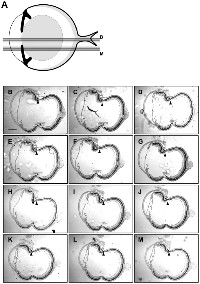
Figure 2. Retinal folding collected in 60 sections.
Schematic of a mouse eye (A), showing the cutting pattern of the serial sections displayed from B-M. Five-micron sections were collected at regular intervals starting at the optic nerve head and continuing outwards relative the center. Approximately 60 sections were collected; every fifth section is shown here. Arrowheads indicate the position of a fold seen in all 60 sections. Retinas used in this experiment are from mice exposed for 24 hours to 1500 mg VGB/kg and exposed to a constant light source (B-M). Scale bar: 200 µm.

