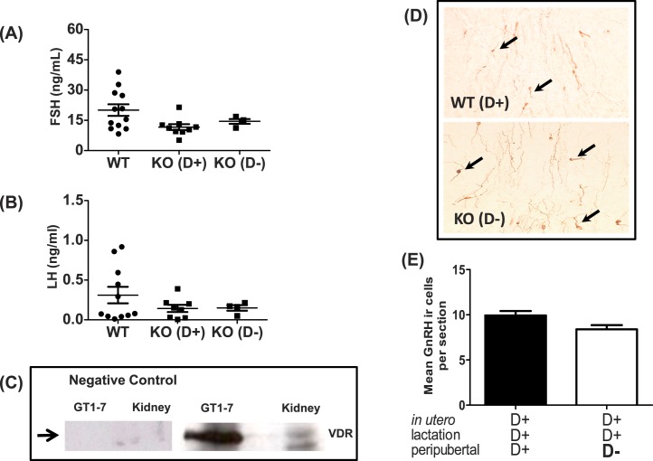
FIG. 7. .
Effects of vitamin D3 on serum gonadotropin levels during mice in diestrus, VDR expression in GT1-7, and density of hypothalamic GnRH neurons. A) Serum FSH in reproduction-aged WT and Cyp27b1 null mice fed a vitamin D3-sufficient diet (KO [D+]) or a D3-deficient diet (KO [D−]) (n = 4–12). B) Serum LH in reproduction-aged WT and Cyp27b1 null mice fed a vitamin D3-sufficient diet (KO [D+]) or D3-deficient diet (KO [D−]) (n = 4–12). C) Western blot showing VDR in cell lysates from GT1-7 neurons and kidney. The positive control is WT mouse kidney, and negative control is GT1-7 and kidney lysate without primary VDR antibody. D) Representative sections of single-label immunohistochemistry (original magnification ×40) showing GnRH neurons (brown cytoplasm) in WT Vit D3+ and Cyp27b1 null Vit D3− mice. Arrows indicate GnRH immunoreactive neurons (n = 4). E) Average number of GnRH neuron numbers per hypothalamic section (means ± SEM) in WT Vit D3+ and Cyp27b1 null Vit D3− mice. Hypothalamic sections reviewed corresponded to plates 25–32 of the Paxinos and Watson mouse atlas [49] and the hypothalamic region between the organum vasculosum of lamina terminalis and the medial POA (n = 4).

