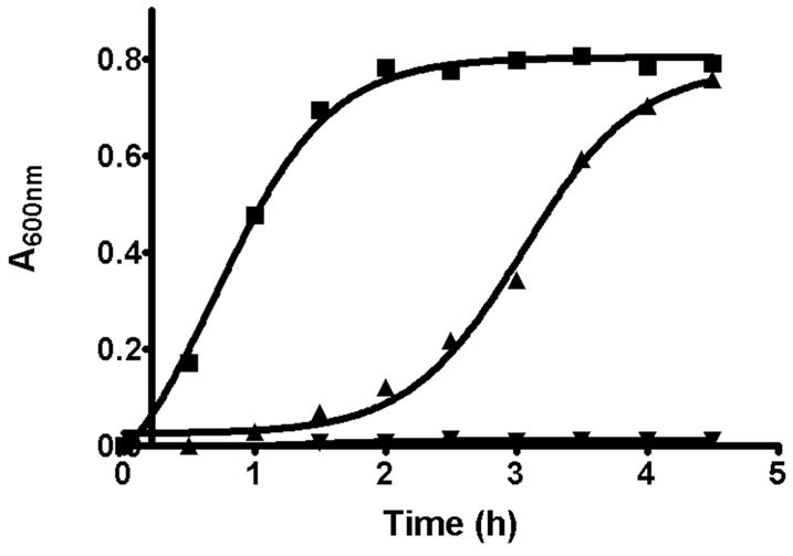
Figure 1.

Recovery of E. faecalis cellular growth after removal of hymeglusin. E. faecalis cells were grown in LB media for 3 hours at 37°C in the presence, or absence, of 25 μM hymeglusin. Cells were pelleted then resuspended in fresh LB media and again incubated at 37°C. Absorbance was measured at 600 nm. The traces correspond to: LB prior to cell pelleting followed by post-resuspension growth in LB (■); hymeglusin/LB followed by post-resuspension LB (▲); hymeglusin/LB followed by post-resuspension hymeglusin/LB (▼). The individual time points are averages of data from three experiments performed in parallel. Curves represent nonlinear regression fits of the data to a sigmoidal equation (GraphPad Prism 4). Inflection times and error estimates are provided in Table 2.
