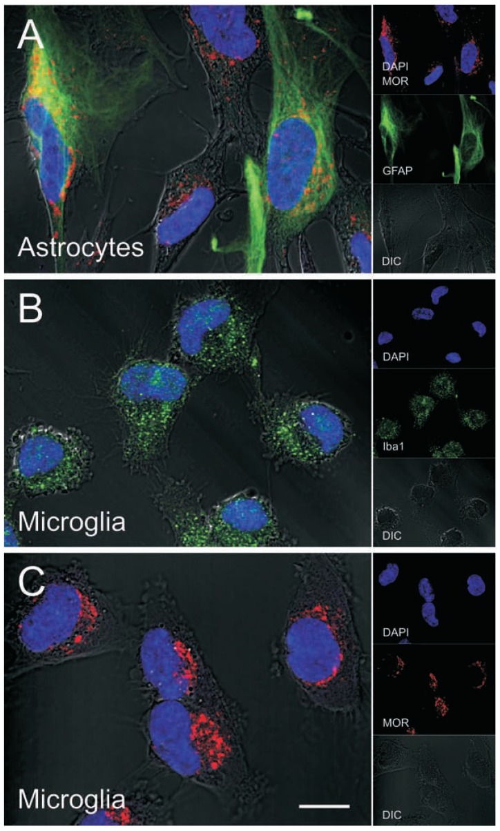
Fig. (3).
MOR immunofluorescence in human astrocytes and microglia. MOR and GFAP co-localization in subsets of primary human astrocytes (A). Astrocytes (catalog number 1800) were obtained from ScienCell Research Laboratories and cultured for 7-10 days according to the manufacturer's instructions. Iba-1 (B) and MOR (C) immunofluorescence in subpopulations of primary human microglia (ScienCell; catalog number 1900-f1) cultured as described for astrocytes. Cells were fixed with 3.7% paraformaldehyde, permeabilized with 0.5% Triton X-100, immuno-labeled, nuclei were stained with DAPI (blue), and images were enhanced by differential interference contrast (DIC) optics. Primary antibodies used were MOR (epitope within amino acids 1-15 of the N-terminus of human MOR) (Novus Biologicals, catalog number NBP1-31180), GFAP (Millipore, catalog number MAB360), and Iba-1 (Wako, catalog number 019-19741); all at a 1:200 dilution. Images were acquired using a Zeiss LSM 700 laser scanning confocal microscope at 63x (1.42 NA) magnification and ZEN 2010 software (Carl Zeiss Inc, Thornwood, NY), and edited using ZEN 2009 Light Edition (Zeiss) and Adobe Photoshop CS3 Extended 10.0 software (Adobe Systems, Inc.).

