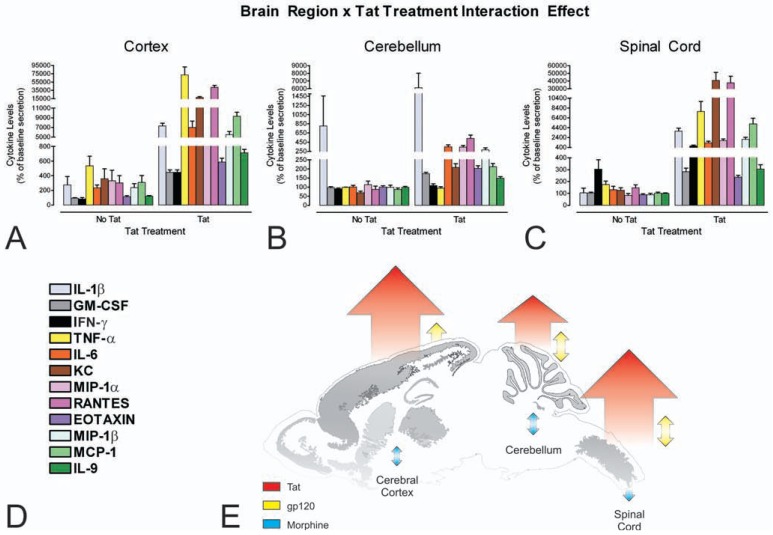
Fig. (4).
Astroglia isolated from the cerebral cortex (A), cerebellum (B), and spinal cord (C) display significant regional differences in the pattern of cytokine release in response to HIV-1 Tat in vitro (A-C). Interestingly, the pattern of cytokine release in response to Tat paralleled the incidence of HIV-1-related neuropathology in the brain and spinal cord (see reference [86]). Striatal astrocytes, analyzed as part of another study, show a far more dramatic interaction between HIV-1 Tat and morphine [72]. Cytokines and chemokines were analyzed simultaneously by multiplex suspension array assays [86]; legend provided in (D). Overall responses to HIV-1 Tat, gp120 and morphine across brain regions are summarized (E) (see text and reference [86] for further explanation). Reprinted with permission from reference [86]. Copyright 2010 American Chemical Society.

