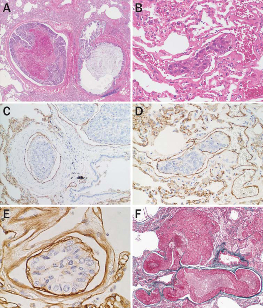
Fig. 3.
Pulmonary tumor emboli of HCC. (a) Large tumor mass composed of multiple tumor nests, and with central necrosis, are embolized in a pulmonary artery (hematoxylineosin stain, original magnification 20×). (b) A single tumor nest is arrested in an arteriole (hematoxylin-eosin stain, original magnification 200×). (c, d) Immunostaining of CD31 highlights tumor-associated blood vessels surrounding tumor emboli both in the pulmonary artery (c) and arterioles (d) (original magnification (c) 100×; (d) 200×). (e) The embolus conserves the trabecular architecture with the basement membrane (Immunostaining of laminin, original magnification 200×). (f) Intravascular growth and extravasation of HCC cells embolized in pulmonary artery (Elastica Masson stain, original magnification 20×)

