FIGURE 5.
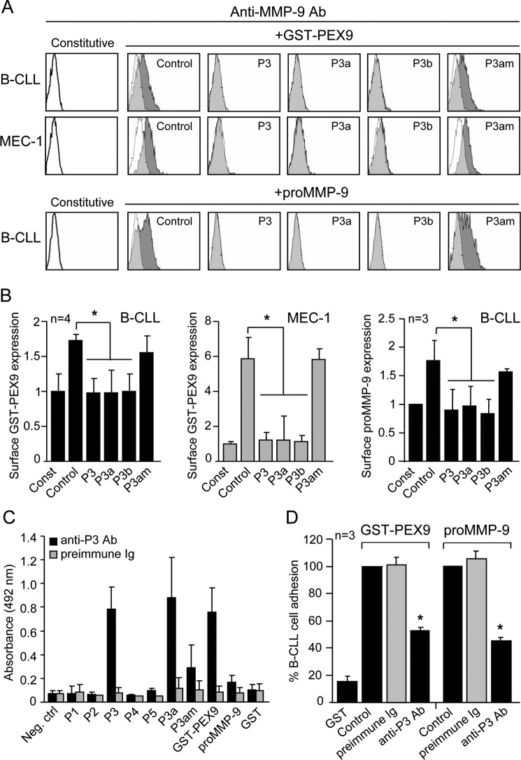
Further effects of P3, P3-derived peptides, and anti-P3 antibodies on B-CLL cells. A, primary B-CLL or MEC-1 cells treated or not for 30 min with the indicated peptides (at the same μm concentrations used in Fig. 4), were incubated with soluble GST-PEX9 (0.4 and 0.6 μm, respectively) or proMMP-9 (60 nm). After 30 min, binding was analyzed by flow cytometry using an anti-MMP-9 antibody. A representative experiment (patient 18 for B-CLL) is shown. White areas represent constitutive (pro)MMP-9 expression; gray areas, (pro)MMP-9 expression upon GST-PEX9 binding. Binding of soluble proMMP-9 to MEC-1 cells was also inhibited by the P3/P3a/P3b peptides and is not shown. B, quantitative values of the soluble binding assays represent the average of four (patients 8, 14, 17, 18; GST-PEX9) or three (patients 14, 17, 18; proMMP-9) B-CLL samples or the average of three different experiments with triplicate determinations (MEC-1). C, the specificity of the anti-P3 antibody was determined by ELISA assays with the indicated immobilized peptides (8 μm) or proteins (2 μm). Negative control (Neg. ctrl), average absorbance of peptides or proteins with only secondary antibody. D, BCECF-AM-labeled B-CLL cells (patients 15, 18, 20) with or without previous incubation with preimmune Ig or anti-P3 antibody were added to wells coated with GST-PEX-9 or proMMP9 and adhesion measured as explained. *, p ≤ 0.05.

