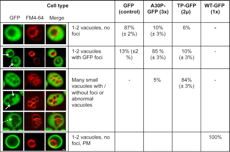
FIGURE 3.
Vacuole morphology and GFP localization in yeast cells expressing non-aggregated α-synuclein-GFP variants. Live-cell fluorescence microscopy of yeast cells (W303) expressing GFP (control), A30P-GFP (3× integrated in the genome), TP-GFP (2-μm plasmid), and WT-GFP (1× integrated in the genome). GAL1-driven α-synuclein-GFP protein expression was induced in galactose-containing medium for 24 h. Yeast vacuoles were stained with FM4-64 and examined for morphology. White arrows point at GFP-foci inside the vacuole. The dashed arrow indicates tubular invaginations of the yeast vacuole. The quantification of different cell types represents an average of three independent experiments. PM, plasma membrane localization of GFP signal. Scale bar = 1 μm.

