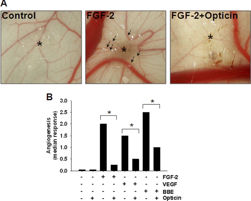
FIGURE 1.
Effects of opticin in CAM ex vivo model. A, representative images showing the effects of a pellet without FGF-2 or opticin (left image), and pellets containing FGF-2 with or without opticin. Asterisk indicates the site of application of the pellet. Under FGF-2 stimulation (middle image), newly formed vessels converge toward where the growth factor-containing pellet was placed (arrows). B, pellets containing growth factors i.e. FGF-2, VEGF165, and BBE, with or without opticin, were applied on the CAM, and the angiogenic response was quantified at day 10. Addition of opticin in combination with FGF-2 (n = 8), VEGF165 (n = 12), and BBE (n = 12) significantly reduced their angiogenic drive on the CAM (*, p = 0.01, p = 0.04, and p < 0.035 for FGF-2, VEGF165, and BBE, respectively).

