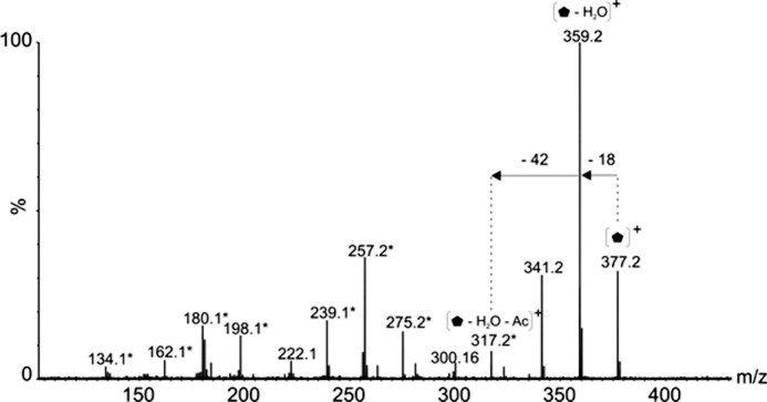
FIGURE 7.

Glycan structural characterization by mass spectrometry. Tandem mass spectrum of singly charged ion at m/z 377 following feCID of intact flagellin protein showed consecutive losses of water and an acetyl group from the parent ion giving rise to an ion at m/z 317. Subsequent losses from this ion gave rise to a fragmentation pattern similar to that observed with nonulosonic acid sugars. Daughters ions marked with an asterisk denote those fragment ions found in MS/MS spectra of pseudaminic acid.  , 376-Da sugar; *, fragment ions common to pseudaminic acid; Ac, acetyl group.
