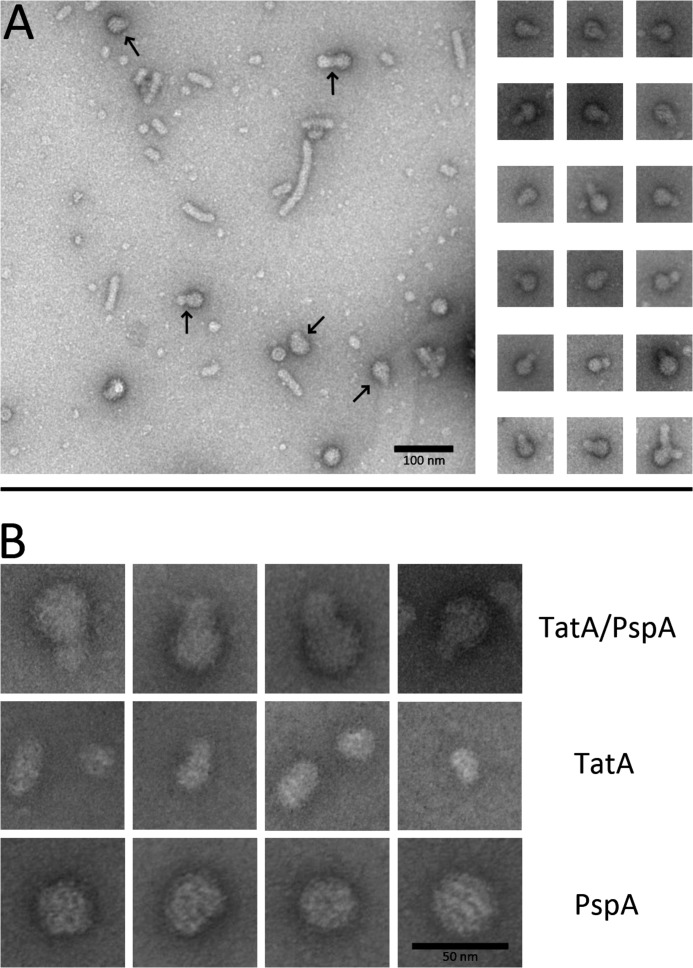
FIGURE 2.
Detection of the TatA-PspA interaction by electron microscopy. A, overview micrograph of negatively stained affinity-purified Strep-tagged TatA, with co-purified PspA (left) and depicted TatA-PspA complexes (right). In the overview image, arrows point to readily recognizable conjunctions of TatA-PspA complexes. Note that longer filament-like TatA stacks, although frequently observed in the overview, were only rarely seen to interact with PspA. B, higher resolution micrographs of TatA-PspA complexes (upper panel), TatA as purified in the absence of PspA (middle panel), and a TatA-free PspA preparation (lower panel). The smaller ∼16 nm diameter TatA particles are seen as predominant interaction partners of PspA and are therefore depicted in the TatA panel of B.

