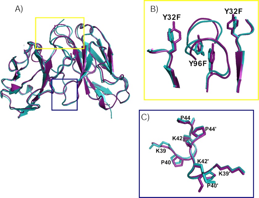
FIGURE 5.
A, superposition of AL-103 2YF (teal) and AL-103 (purple) crystal structures shows few structural differences. B (yellow box), detailed view of the Tyr-to-Phe mutations shows rotation of Phe-32 (from residues 30–36 and 91–96). C (blue box), close up of alterations in loops between β-strands C and C′ (residues 39–44) show minor differences.

