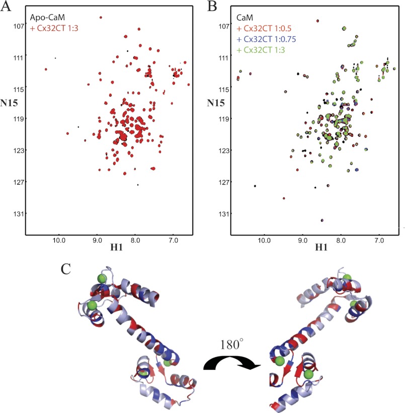
FIGURE 6.
NMR studies characterizing the CaM residues affected upon Cx32CT binding. A, overlaid 15N-HSQC spectrum of apo-CaM (black) with apo-CaM-Cx32CT (red). B, change in chemical shift position on CaM binding to Cx32CT in the presence of 5 mm Ca2+. 15N-HSQC titration of 15N-CaM with unlabeled Cx32CT. Each titration contained the same concentration of 15N-CaM (70 μm) with different concentrations of the Cx32CT. The cross-peak color changes according to the concentration ratio (CaM:Cx32CT 1:0 (black), 1:0.5 (red), 1:0.75 (blue), 1:3 (green)). C, three-dimensional representation of perturbed residues in Ca2+-CaM (Protein Data Bank code 3CLN) upon binding to Cx32CT. Residues with amide proton resonance intensity changes over 10% are shown in blue (weaker interaction) and when peaks broaden beyond detection shown in red (stronger interaction). Calcium ions are shown in green.

