
FIGURE 2.
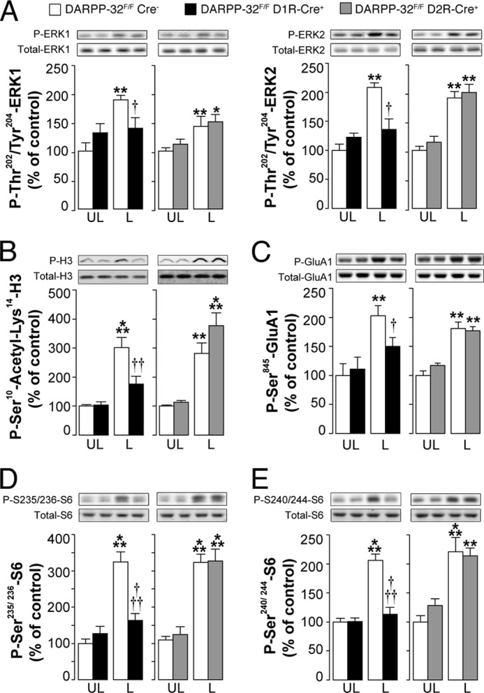
Selective deletion of DARPP-32 in striatonigral MSNs decreases l-DOPA-induced phosphorylation of ERK, histone H3, Glu-1A, and rpS6. DARPP-32F/FD1RCre+ mice, DARPP-32F/FD2RCre+ mice, and DARPP-32F/FCre− littermates were lesioned unilaterally with 6-OHDA, treated for 10 days with 10 mg/kg of l-DOPA, and killed 30 min after the last injection. Top rows show representative autoradiograms obtained using antibodies against total or phosphorylated ERK (A), histone H3 (B), Glu-1A (C), and rpS6 (D and E). Bottom rows are summary of results showing means ± S.E. (n = 6). *, p < 0.05; **, p < 0.01; ***, p < 0.001 versus unlesioned (UL) DARPP-32F/FCre− and DARPP-32F/FD2RCre+ mice treated with l-DOPA; †, p < 0.05; ††, p < 0.01; †††, p < 0.001 versus 6-OHDA lesioned DARPP-32F/FCre− mice treated with l-DOPA (L); two-way ANOVA, followed by Bonferroni-Dunn test. A significant interaction between treatment and genotype was found in DARPP-32F/FD1RCre+ mice (F(1, 20) = 8.9, p < 0.01 for phospho-ERK1; F(1, 20) = 11.47, p < 0.01 for phospho-ERK2; F(1, 22) = 8.32, p < 0.01 for phospho-Ser10-acetyl-Lys-14-histone H3; F(1, 20) = 7.44, p < 0.05 for phospho-Ser-845-Glu-1A; F(1, 20) = 7.64, p < 0.05 for phospho-Ser-235/236-rpS6; F(1, 20) = 7.74, p < 0.05 for phospho-Ser-240/244-rpS6). A significant effect of the treatment was found in DARPP-32F/FD2RCre+ mice (F(1, 20) = 26.64, p < 0.001 for phospho-ERK1; F(1, 20) = 30.95, p < 0.001 for phospho-ERK2; F(1, 22) = 36.98, p < 0.001 for phospho-Ser-10-acetyl-Lys-14-histone H3; F(1, 20) = 35.44, p < 0.001 for phospho-Ser-845-Glu-1A; F(1, 20) = 80.3, p < 0.001 for phospho-Ser-235/236-rpS6; F(1, 20) = 44.41, p < 0.001 for phospho-Ser-240/244-rpS6).