FIGURE 2.
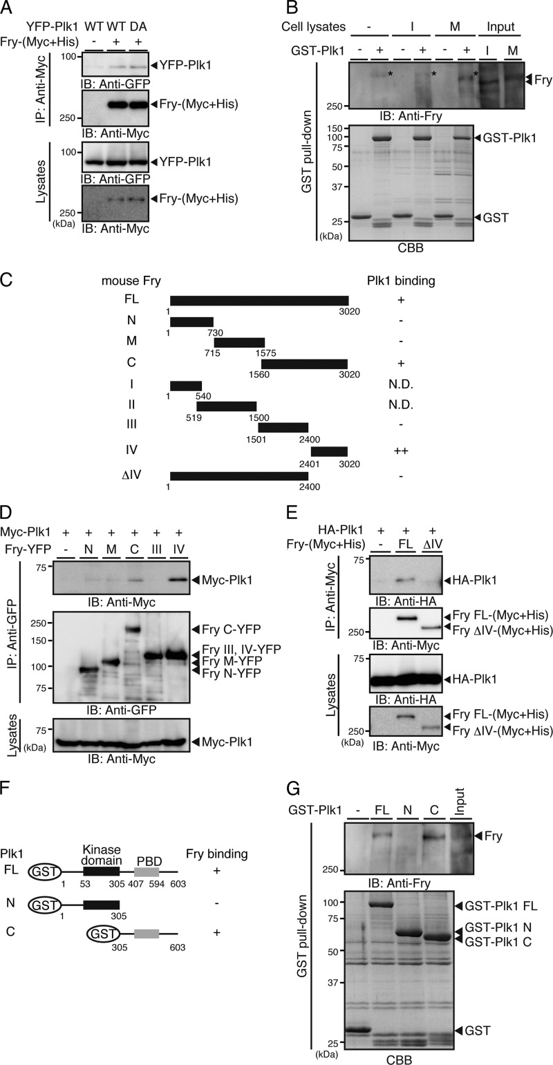
Fry binds to the PBD of Plk1. A, Fry binds to Plk1. YFP-Plk1(WT or DA) and Fry-(Myc+His) were coexpressed in 293T cells. Cell lysates were immunoprecipitated (IP) with anti-Myc antibody and analyzed by anti-Myc and anti-GFP immunoblotting (IB). B, mitotic Fry binds to Plk1. HeLa cells were synchronized in interphase (I) by double thymidine block or in early mitotic phase (M) by nocodazole exposure. Lysates were incubated with GST or GST-Plk1-immobilized beads, and the bound proteins were immunoblotted with anti-Fry antibody. The asterisk indicates the nonspecific band that was also detected in the lane of GST-Plk1 beads without cell lysates. C, structure of Fry deletion mutants. D and E, mapping of the Plk1-binding region of Fry. Fry deletion mutants and Plk1 were coexpressed in 293T cells. Lysates were immunoprecipitated with anti-GFP or anti-Myc antibody and analyzed by immunoblotting. F, structure of Plk1 deletion mutants. G, mapping of the Fry-binding region of Plk1. Lysates of nocodazole-arrested HeLa cells were subjected to GST pull-down assays using GST-Plk1 or Plk1 fragments. Precipitates were analyzed by immunoblotting with anti-Fry antibody.

