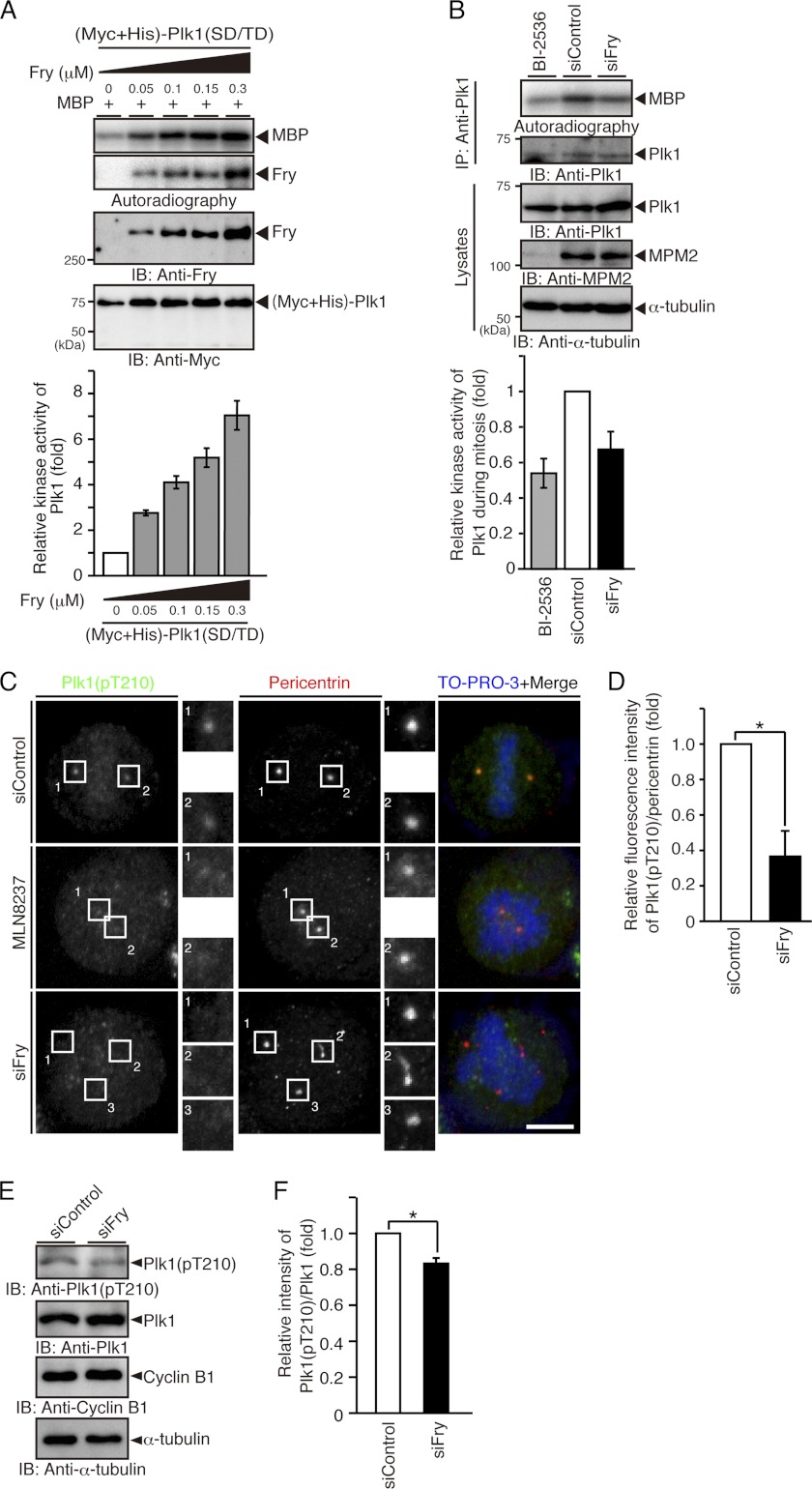
FIGURE 5.
Fry depletion decreases the kinase activity and the Thr-210 phosphorylation level of Plk1. A, Fry increases the kinase activity of Plk1 in a concentration-dependent manner. Purified (Myc+His)-Plk1(SD/TD) was mixed with recombinant Fry and subjected to in vitro kinase assay using MBP as a substrate. The relative kinase activity of Plk1 is shown as the means ± S.E. of triplicate experiments. IB, immunoblotting. B, Fry depletion reduces the kinase activity of mitotic Plk1. HeLa cells transfected with siRNAs were synchronized in early mitosis with thymidine-nocodazole. Mitotic HeLa cells were collected by the mechanical shake-off method. Endogenous Plk1 was immunoprecipitated (IP) and subjected to an in vitro kinase assay using MBP as a substrate. Mitotic HeLa cells exposed to BI-2536 (1 μm) were used as a background control. The relative kinase activity of Plk1 is shown as the means ± S.E. of triplicate experiments. C, Fry depletion decreases the level of Thr-210 phosphorylation of Plk1 on spindle poles. HeLa cells transfected with siRNAs were cultured in growth medium for 12 h and in thymidine-containing medium for 36 h. They were then released from thymidine arrest for 12 h before being fixed and stained with anti-Plk1 pT210 (green) and anti-pericentrin (red) antibodies. DNA was stained with TO-PRO-3 (blue). For Aurora A inhibition, after release from thymidine block for 10 h, HeLa cells transfected with control siRNA were incubated for 2 h in medium containing MLN8237 (100 nm) and MG132 (10 μm). Magnified images of the white boxes are also shown. Scale bar, 5 μm. D, relative fluorescence intensity of Plk1(pT210) normalized to the intensity of pericentrin on spindle poles. HeLa cells were treated as in C. The fluorescence intensity in a 2-μm-diameter circular region surrounding the spindle pole was measured. The relative fluorescence intensity is shown as the means ± S.E. of triplicate experiments. *, p < 0.05. E, immunoblot analysis of the level of Thr-210 phosphorylation of Plk1. HeLa cells transfected with siRNAs were synchronized at early mitotic phase with thymidine-nocodazole treatment and collected by the mechanical shake-off procedure. Cell lysates were analyzed by immunoblotting with indicated antibodies. The relative intensity of Plk1(pT210) immunoblot normalized to the intensity of Plk1 is shown as the means ± S.E. of triplicate experiments. *, p < 0.05.

