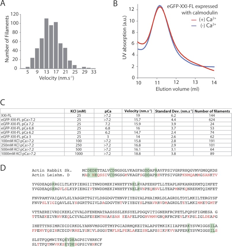
FIGURE 7.
Gliding filament assay. A, the distribution of velocities of rhodamine-phalloidin-labeled actin filaments gliding over eGFP-FL-XXI. Myosin was deposited on nitrocellulose surfaces at 200 μg/ml. B, SEC (Superdex-200) of FL-XXI coexpressed with calmodulin in the absence and presence of calcium pCa 4.1. Experiments were repeated a minimum of three times from three separate protein purifications. C, comparison of the mean velocity obtained with myosin-XXI at different ionic strengths and calcium concentrations. All experiments were carried out at 22 °C. Data were collected from at least three separate flow cells for each condition. D, multiple sequence alignment of rabbit skeletal and L. donovani actin. Differences are marked in red. Residues on the surface of actin involved in myosin binding according to Kabsch et al. (35) are highlighted in green.

