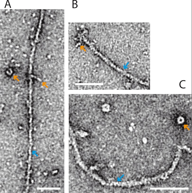
FIGURE 8.
Electron microscopy. Negative stain electron microscopy of eGFP-FL-XXI bound to F-actin (see “Experimental Procedures”). A, single myosin-XXI molecules (orange arrow) are bound to an actin filament (blue arrow). When attached to the surface on their own they often adopted a compact structure (orange arrow). B, several myosin-XXI molecules are attached to the end of an actin filament. C, myosin-XXI molecules form a compact, ring-like structure when detached from actin. Scale bars = 50 nm.

