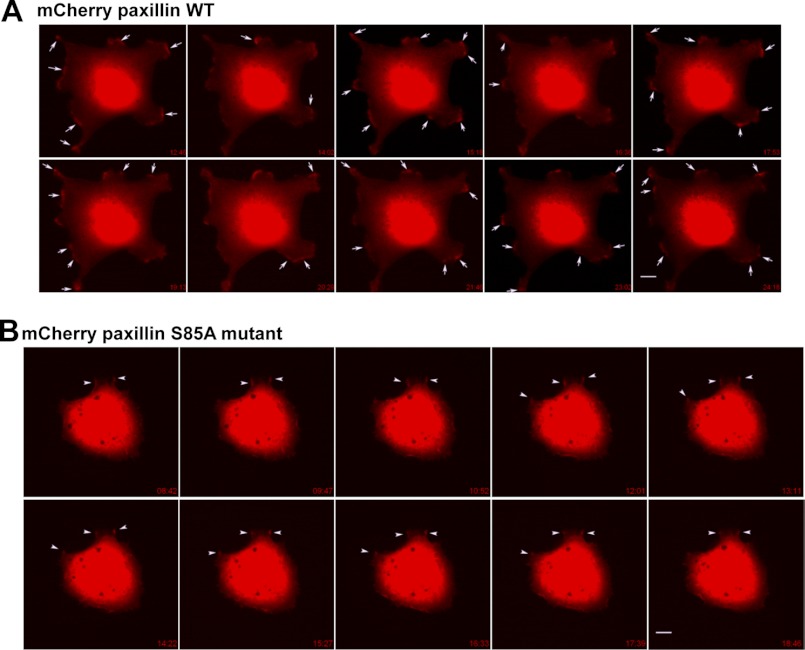
FIGURE 7.
Time-lapse images of mCherry-paxillin (WT or S85A)-expressing cells. Cells were transiently transfected with mCherry-tagged WT (A) or S85A mutant (B) paxillin for 36 h and replated onto collagen I (2 μg/ml)-precoated coverslips for 30 min within 0.2% BSA and DMEM-H before analysis with time-lapse microscopy. Images were saved over 30 min, and snap pictures in series over at least 10 min are represented. A, in images of mCherry-WT paxillin-transfected cells, dynamic turnover of paxillin-positive spots (arrows) is evidenced by newly formed spots compared with the immediately preceding snap picture. B, in images of mCherry-S85A paxillin-transfected cells, the positive staining spots (arrowheads) were less dynamic, and the staining was sustained for longer times. Images shown represent cells from several analyses (mCherry-WT paxillin, n = 9; and mCherry-S85A paxillin, n = 11).

