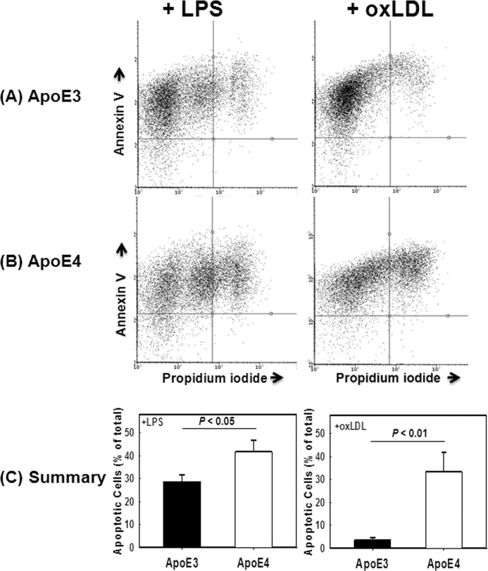
FIGURE 4.
Agonist-induced death of APOE3 and APOE4 macrophages. Peritoneal macrophages from APOE3 (A) and APOE4 (B) mice were incubated with 50 ng/ml LPS (left panels) or 100 μg/ml oxLDL. Cell death was assessed by flow cytometry based on expression of annexin V and propidium iodide. Representative flow cytometry data are shown in A and B. C shows the mean ± S.D. from four separate experiments each performed with cells obtained from at least three mice per group. Differences as noted were evaluated by Student's t test.

