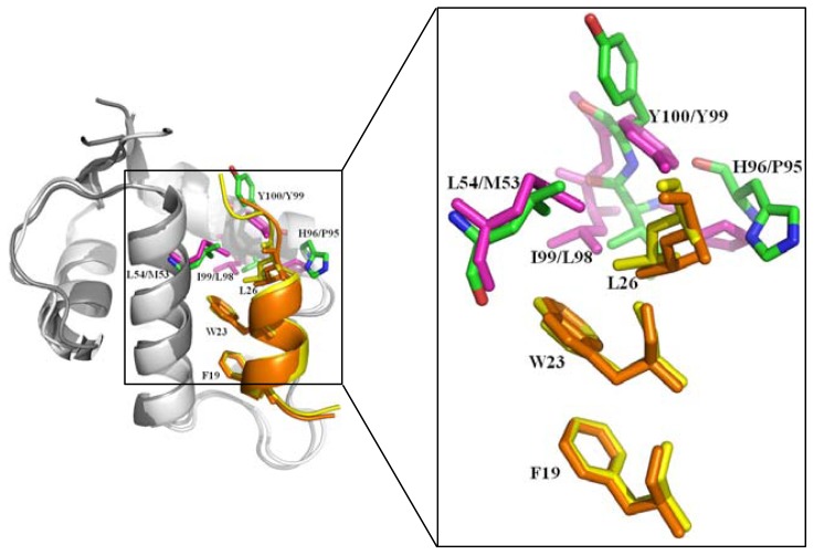
Figure 3.
The superimposition of the structures of p53-MDM2 and p53-MDMX complex. The complex of p53-MDM2 (PDB 1YCR) [18] is shown in cartoon, p53 TAD fragment (residues 17–29) is shown in yellow and the three most important residues Phe19, Trp23 and Leu26 are shown in stick, MDM2 (residues 25–109) is shown in white and the four residues Leu54, His96, Ile99 and Tyr100 in MDM2 are shown in green and stick; The complex of p53-MDMX (PDB 3DAB) [27] is shown in cartoon, p53 TAD fragment (residues 17–29) is shown in orange and the three most important residues Phe19, Trp23 and Leu26 are shown in stick, MDMX (residues 23–110) is shown in grey and the equivalent residues Met53, Pro95, Leu98 and Tyr99 in MDMX are shown in light magenta and stick; the spatial orientation of the side chains of Phe19 and Trp23 of p53 are highly similar, but that of Leu26 is different in p53-MDM2 and p53-MDMX complex. (Insert) Magnified view of important residues of p53 and MDM2/X. Figures were created with Pymol (http://pymol.org) [20].

