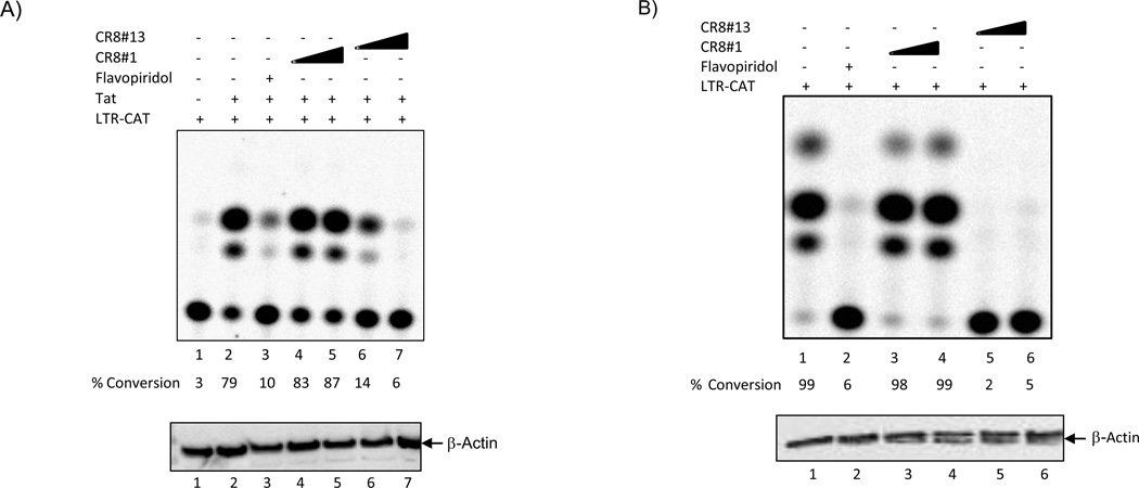
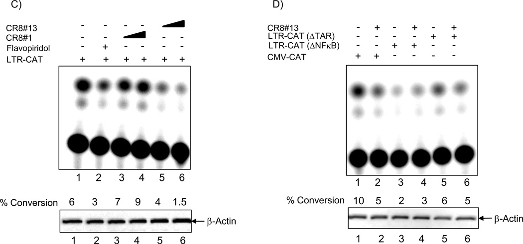
Figure 1. Effect of CR8#13 on the HIV-1 Promoter in Cells.
A) Early to mid log phase CEM cells were electroporated with either LTR-CAT alone (5µg) or with CMV-Tat (Pc Tat; 1.5µg). Cells were processed for CAT assay 48 hours later. Various concentrations of CR8#1 and CR8#13 (50 and 100 nM) were used immediately after plasmid transfection. Flavopiridol (50 nM) was used as a positive control for inhibition of cdk9/T1 in cells. Ten micrograms of total lysate was used in western blot for presence of β-actin. B) TNF-α treated OM10.1 cells were electroporated with LTR-CAT (5µg) and cells were processed for CAT assay 48 hours later. Similarly to Panel A, concentrations of drugs were used immediately after electroporation of cells. C) Same as Panel B, where OM10.1 cells were electroporated with LTR-CAT and processed 48 hours later for CAT assay. These cells were not treated with TNF-α and therefore no Tat should be available for activation of the LTR-CAT construct. D) Same as Panel A, where CEM cells were electroporated with 5 µg of either CMV-CAT, LTR-CAT (ΔNF-κB), or LTR-CAT (ΔTAR; TM26). Samples were treated with CR8#13 (100 nM) immediately after transfection and CAT assay performed 48 hours later. Ten microgram of total extract was used for β-actin western blot. E) Total cell extracts (TNF-α treated OM10.1; 2.5 mg) were used with 25 µg of α-cdk9 antibody for immunoprecipitation. Samples were incubated overnight at 4°C and protein A + G added next day for 2 hours. IPed material was washed twice with TNE150 + 0.1% NP-40 and twice kinase buffer. The IPed material was divided into 6 tubes with histone H1, kinase reaction mix, and drugs were added to each tube. Samples were incubated for 30 minutes/37°C followed by separation on 4–20% SDS/PAGE. Two concentrations of CR8#1 and CR8#13 (10 and 50 nM) were used for kinase reactions. Flavopiridol (10 nM) was used as a control. Counts are from Molecular Dynamics phosphorimager analysis. F) Similar to Panel E, where 10 µg of α-cdk2 and α-cdk4 antibodies were used for immunoprecipitation overnight followed by in vitro kinase assay next day using histone H1 as substrate. A final concentration of 50nM of CR8#13 was used for in vitro assay. The “K” units are in counts of 1000 from the phosphorimager (Molecular Dynamics software). "CE" and "NP" stand for cytosolic extract and nuclear pellet, respectively.


