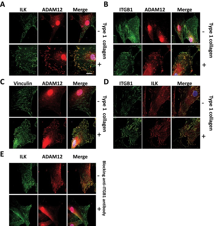
FIGURE 3:
ADAM12L and ILK are recruited to focal adhesion structures upon β1 integrin stimulation. HSCs were cultured on plastic dishes (–) or on dishes coated with type I collagen (+) overnight in medium containing 2% FBS. Cell were immunostained with antibodies against anti-ADAM12, anti-ILK, anti-vinculin, and anti-ITGB1 as indicated, followed by tetramethylrhodamine isothiocyanate– or fluorescein isothiocyanate–labeled secondary IgG. Representative fields are shown. Colocalization results in yellow cellular staining. (A) Localization of ADAM12 and ILK. (B) Localization of β1 integrin (ITGB1) and ADAM12. (C) Localization of vinculin and ADAM12. (D) Localization of ILK and β1 integrin. (E) HSCs were preincubated with anti–β1 integrin blocking antibodies or control mouse IgG1 before seeding on type I collagen. Scale bars, 10 μm.

