
FIGURE 5:
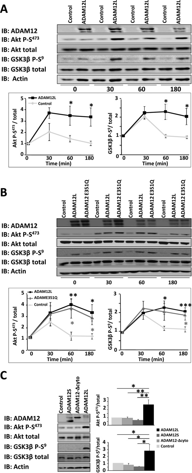
Expression of ADAM12L augments phosphorylation of Akt and GSK-3. Cos7 cells were transfected with empty vector (control) or ADAM12 constructs and cultured for 48 h. The cells were then plated on dishes coated with type I collagen for the indicated times, and the levels of phosphorylated Akt (Akt P-S473), phosphorylated GSK-3β (GSK3β P-S9), total Akt, and GSK-3β were determined by Western blot analysis (IB). Results are expressed as the mean ± SD of three independent experiments (*p < 0.05; **p < 0.01; ***p < 0.01). (A) ADAM12L vs. control. (B) ADAM12L vs. the catalytically deficient ADAM12-E351Q mutant. (C) ADAM12L vs. ADAM12S and ADAM12-Δcyto (time, 60 min).