FIGURE 1:

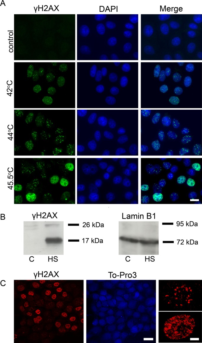
Hyperthermia induces the phosphorylation of histone variant H2AX at Ser-139 in human cells. (A) Immunofluorescence analysis of γH2AX in control (untreated) human mcf-7 cells and cells that were heat-stressed at different temperatures (42, 44, and 45.5°C for 30 min). The DNA was stained with DAPI. Scale bar: 20 μm. (B) Western blot analysis of γH2AX in the nuclei of control (untreated, C) and heat-shocked (45.5°C, 30 min; HS) mcf-7 cells. Lamin B1 was used as a loading control. (C) Immunofluorescence analysis of heat-treated (45.5°C, 30 min) mcf-7 cells performed using a Leica SP2 confocal laser scanning microscope (scale bar: 20 μm). Enlarged cells with two different patterns of γH2AX distribution are shown on the right (scale bar: 5 μm). The DNA was stained with To-Pro 3 iodide fluorescent dye.
