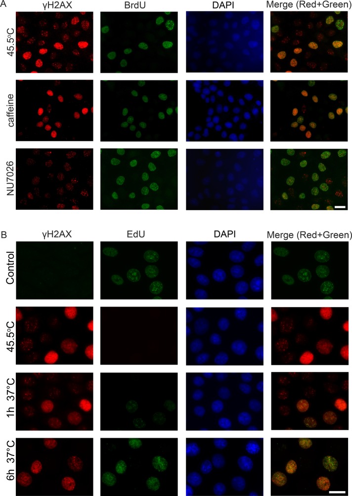
FIGURE 6:
Characterization of HS-induced γH2AX foci in human mcf-7 cells. (A) HS-induced H2AX phosphorylation is mediated by different PIKKs. Human mcf-7 cells were treated with either an ATM/ATR inhibitor (caffeine; 10 mM, 3 h) or a DNA-PKcs inhibitor (NU7026; 10 μM, 6 h), pulse-labeled with BrdU (100 μM, 20 min), heat stressed (45.5°C, 30 min), and double-immunostained against γH2AX (red) and BrdU (green). The DNA was stained with DAPI (blue). A superimposition of the green and red channels is shown as the “Merge.” Scale bar: 20 μm. (B) The kinetics of γH2AX focus recovery after HS in mcf-7 cells. Human mcf-7 cells that were either untreated, treated with HS (45.5°C, 30 min), or treated with HS and allowed to recover for the indicated time intervals (1 and 6 h) were pulse-labeled with EdU (10 μM, 30 min) and then immunostained against γH2AX (red). EdU (green) was revealed by Click Chemistry. The DNA was stained with DAPI (blue). A superimposition of the green and red channels is shown as the “Merge.” Scale bars: 20 μm.

