FIGURE 2:
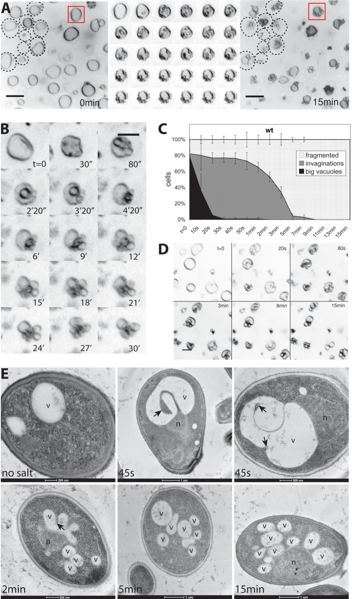
Remodeling of invaginated vacuoles into vesicles. Vacuole fragmentation was induced in FM4-64–stained wild-type (BJ3505) cells by addition of 0.5 M NaCl. (A) Vacuole structure of a group of cells before (left) and 15 min after salt addition (right). The cell perimeters for some cells are indicted by dashed lines. Middle, a magnified view of the vacuole marked by a red square. Images of this vacuole were taken every 20 s during the 10 min after salt addition. Scale bars, 2 μM. (B) Maximum-intensity projection of optical sections of a single vacuole during fragmentation, covering 4 μM in the z-direction. (C) Quantification of vacuole fragmentation in BJ3505 in vivo. Cells were scored according to their vacuolar morphology at different time points after salt addition. We distinguished cells with big intact vacuoles without invaginations, cells with vacuolar invaginations of various sizes, and cells with mainly fragmented vacuoles. Three independent image series were analyzed for each time point. (D) Vacuolar fission in CRY1 wild-type cells, a protease-competent strain with vacuoles of similar size as BJ3505. Cells were stained with FM4-64 and imaged at the indicated times after salt addition. (E) Electron micrographs of BJ3505 cells subjected to high-pressure freezing and freeze substitution at different time points after salt shock. Arrows mark invaginations. v, vacuole; n, nucleus.

