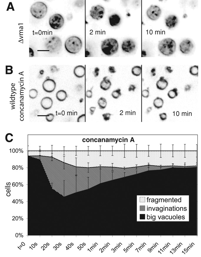
FIGURE 4:

Necessity of the vacuolar proton gradient for vacuole invagination. Cells were stained with FM4-64 and imaged at the indicated time points after addition of 0.5 M NaCl. (A) A Δvma1 strain. (B) Wild-type (BJ3505) cells treated with concanamycin A for 60 min. (C) Quantification of morphological changes over time for vacuoles of concanamycin A–treated wild-type cells. Compare with the graph for nontreated cells in Figure 2C.
