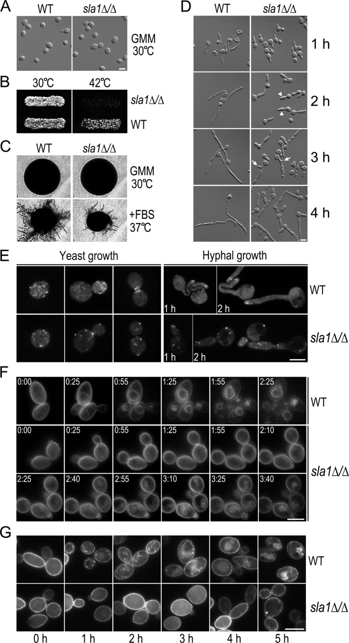
FIGURE 3:
Defects of the sla1∆/∆ mutant. (A) Morphology of sla1Δ/Δ yeast cells. WT (BWP17) and sla1∆/∆ (GZY602) cells were grown in GMM at 30°C. Bar, 5 μm, in this and subsequent figures. (B) Temperature sensitivity of sla1Δ/Δ cells. WT and sla1Δ/Δ cells were grown on a GMM plate at 30°C and replicated to a new GMM plate for incubation at 42°C for 24 h. (C) Defective hyphal growth of sla1Δ/Δ cells on solid medium. WT and sla1∆/∆ cells were streaked on either a GMM plate at 30°C or a GMM plate containing 20% FBS at 37°C. After incubation for 24 h, single colonies were photographed. (D) Defective hyphal growth of sla1Δ/Δ cells in liquid medium. WT and sla1Δ/Δ yeast cells were induced for hyphal growth in YPD containing 20% of FBS at 37°C. Arrowheads and arrows indicate swelling hyphae and buds in subapical cells, respectively. (E) Actin cytoskeleton abnormality in sla1Δ/Δ cells. WT and sla1∆/∆ cells were either grown as yeast in YPD at 30°C or induced for hyphal formation in YPD with 20% of FBS at 37°C for 1–2 h. Cells were stained with rhodamine–phalloidin to visualize the actin cytoskeleton under a fluorescence microscope. (F) Defective FM4-64 uptake in sla1Δ/Δ cells. Fresh WT and sla1Δ/Δ yeast cells were incubated with 20 μM of FM4-64 at 30°C for 10 min before time-lapse microscopic analysis. Representative images taken at the indicated time points (h:min) are shown. (G) Delayed Arn1-GFP internalization in sla1Δ/Δ cells. WT (GZY746) and sla1∆/∆ cells (GZY747) expressing Arn1-GFP were grown in GMM at 30°C and then incubated with 200 μM FC to initiate Arn1-GFP internalization. Samples were taken at 1-h intervals for microscopy.

