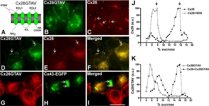
FIGURE 2:
Cx26GTAV formed gap junction plaques when coexpressed with wild-type Cx26 but not when expressed alone or in combination with wild-type Cx43. (A) Diagram of the Cx26GTAV mutant. In this mutant, amino acids 37–40 from Cx26 were substituted for the corresponding sequence from Cx43, GTAV; a hemagglutinin epitope (HA) was appended to the carboxy terminus of the protein. The location of the GTAV sequence at the extracellular end of the first TM domain (TM1) is indicated. ECL1 and ECL2, first and second extracellular loops; ICL, intracellular loop; NH2 and COOH, amino and carboxyl termini. (B and C) Cellular distributions of Cx26GTAV (B) and wild-type Cx26 (C) in HeLa cells stably transfected with Cx26GTAV (HeLaCx26GTAV) or wild-type Cx26 (HeLaCx26) subjected to immunofluorescence using anti-HA (B) or anti-Cx26 (C) antibodies. (D–F) Immunolocalization of Cx26GTAV (D) and wild-type Cx26 (E) in HeLaCx26 transiently transfected with Cx26GTAV after double immunofluorescence using anti-HA (D) and anti-Cx26 (E) antibodies. The merged image is shown in (F). (G–I) Images of HeLaCx26GTAV transiently transfected with wild-type Cx43EGFP showing immunolocalization of Cx26GTAV (G) using anti-HA antibodies and GFP fluorescence localization of wild-type Cx43-EGFP (H). The merged image is shown in (I). Gap junction plaques are indicated by arrows. Scale bar: 15 μm. (J) Graphs represent the levels of Cx26 in sucrose gradient fractions detected by immunoblotting using anti-Cx26 antibodies. Triton X-100–soluble extracts from HeLaCx26 cells treated with SDS (open circles) or left untreated (closed circles) were subjected to sedimentation velocity through 5–20% sucrose gradients. (K) Graphs represent the levels of Cx26GTAV detected by immunoblotting of sucrose gradient fractions using anti-HA antibodies. Triton X-100–soluble material from HeLaCx26GTAV cells (open squares) or HeLaCx26 cells transiently transfected with Cx26GTAV (closed squares) was subjected to sedimentation velocity through 5–20% sucrose gradients. Levels of connexin are presented in arbitrary units (a.u.) after densitometric analysis of the respective immunoblots. Arrows indicate the percentage of sucrose at which Cx26 monomers and hexamers sediment.

