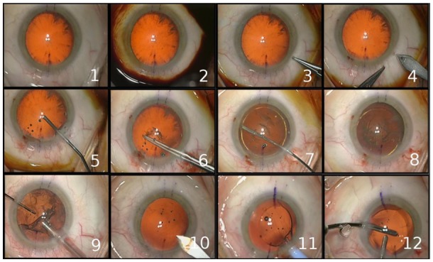
Fig. 6.

Typical digital microscope frames for the 12 surgical phases: 1-preparation, 2-betadine injection, 3-lateral corneal incision, 4-principal corneal incision, 5-viscoelastic injection, 6-capsulorhexis, 7-phacoemulsification, 8-cortical aspiration of the big pieces of the lens, 9- cortical aspiration of the remanescent lens, 10-expansion of the principal incision, 11-implantation of the artificial IOL, 12- adjustment of the IOL+ wound sealing
