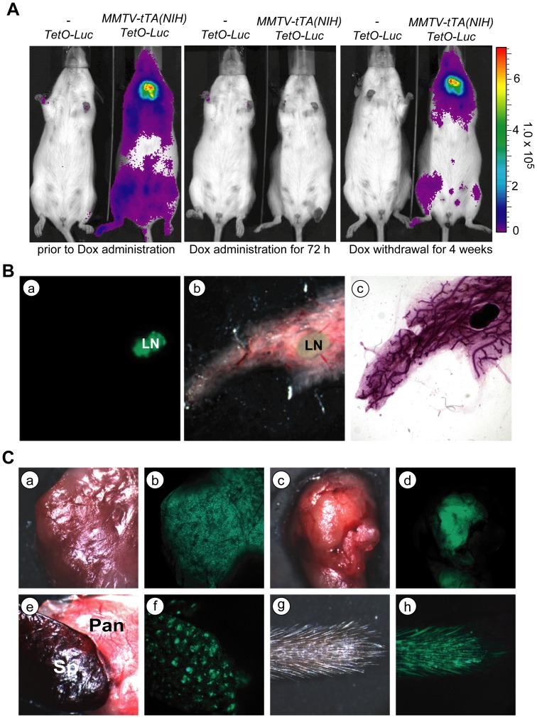
Figure 1. The MMTV-tTA(NIH) transgenic strain shows a strong tTA-mediated transactivation in many tissues.
A. Bioluminescence imaging on a MMTV-tTA(NIH)/TetO-Luc double transgenic female and a TetO-Luc single transgenic littermate negative control. The three panels show the same animals prior to and 3 days after treatment with Dox as well as 33 days after Dox withdrawal. B. Fluorescence images of the GFP reporter expression in the mammary gland of a MMTV-tTA(NIH)/TetO-H2B-GFP double transgenic female. Panels a. and b. show GFP and bright-field images of an unfixed mammary gland. Panel c. shows the same gland after fixation and carmine red staining for visualizing mammary ducts. LN: lymph node. C. GFP expression in a variety of organs from a MMTV-tTA(NIH)/TetO-H2B-GFP double transgenic female; a, b: salivary gland; c, d: thymus; e, f: spleen (Sp) and pancreas (Pan); g, h: tail.

