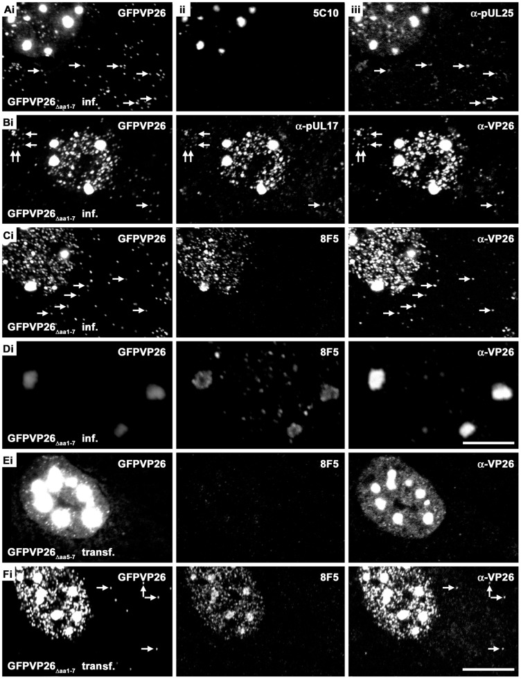
Figure 5. Nuclear aggregates contain capsid proteins.
Vero cells were infected (inf.) with 10 PFU/cell of HSV1(17+)blueLox-GFPVP26Δaa1–7 for 9 h (A–D) or transfected (transf.) with pHSV1(17+)blueLox-GFPVP26Δaa5–7 (E) or pHSV1(17+)blueLox-GFPVP26Δaa1–7 (F) for 24 h. The cells were fixed with PFA and permeabilized with TX-100. GFPVP26 was detected by its intrinsic fluorescence (left column). Furthermore, the cells were labeled with different VP5 antibodies (MAb 8F5 or 5C10) and with α-VP26, α-pUL25 or α-pUL17, and analyzed by confocal fluorescence microscopy. For row D, the specimen was scanned at higher magnification and lower photomultiplier settings. The arrows point to cytoplasmic capsids. Bars: 5 µm (D), 10 µm (F).

