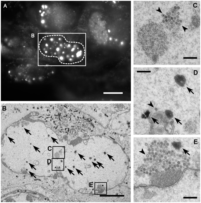
Figure 6. Nuclear aggregates do not contain capsids.
RPE cells were infected with HSV1(KOS)-GFPVP26Δaa1–7 at an MOI of 10 PFU/cell. At 19.5 h, the cells were analyzed by fluorescence microscopy to identify nuclei with bright fluorescence spots (A). After fixation, embedding and sectioning these cells were further analyzed by electron microscopy (B–E). The nuclei contain nuclear capsids (arrowheads) as well as the amorphous electron dense material representing the aggregates (arrows) identified by fluorescence microscopy. The aggregates do not contain capsids (arrowheads). Bars: 10 µm (A), 5 µm (B), and 500 nm (C–E).

