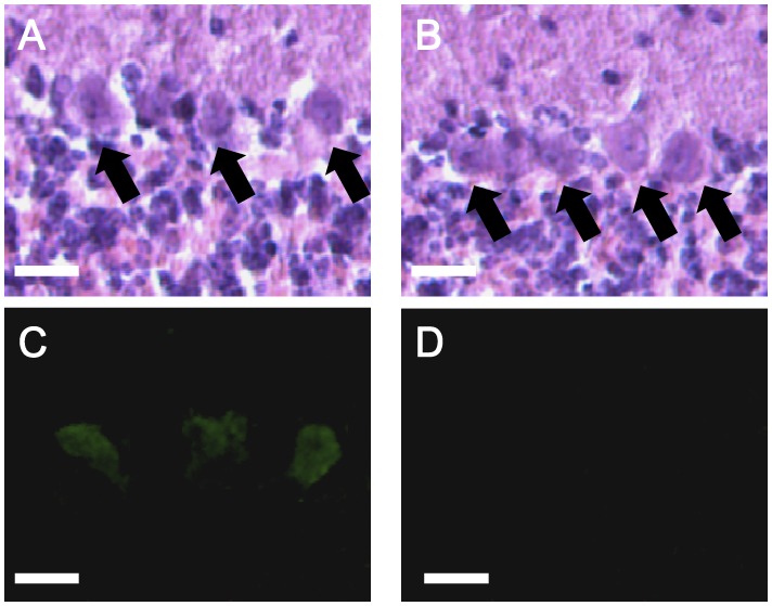
Figure 4. Representative histochemistry in the cerebellum of tottering-6j mice.
Hematoxylin eosin (HE) staining of 6j/6j (A) and +/+ (B) cerebella, and tyrosine hydroxylase (TH) staining of 6j/6j (C) and +/+ (D) cerebella are shown. TH was detected in the Purkinje cells of the 6j/6j mice (n = 6) but not in those of +/+ mice (n = 6). Arrows point to Purkinje cell somata. Scale bar, 20 µm.

