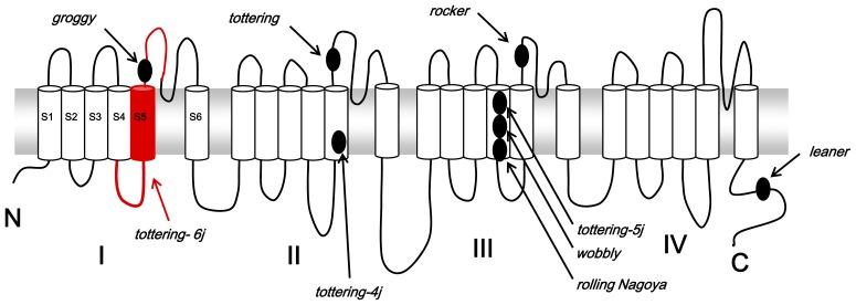
Figure 5. Proposed transmembrane topography of the Cav2.1α1 subunit and positions of known mutations identified in the Cacna1a mutant mice and rat.
The deletion region including part of the S4–S5 linker, S5, and a part of S5–S6 in domain I of Cav2.1α1 in the tottering-6j mice is shown by the red line.

