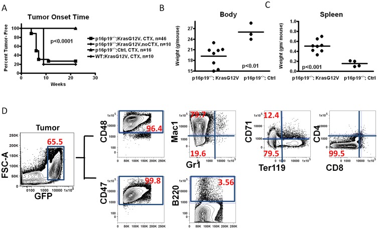
Figure 2. Identical leukemogenic system generates histiocytic sarcoma in the hind limb of NOD.SCID mice when injected into the gastrocnemius muscle.
(A) Kaplan-Meier curve showing the fraction tumor-free mice at the indicated time after transplant of p16p19−/−; Kras(G12V)-GFP with or without cardiotoxin (CTX) pre-injury, or of p16p19−/− ;Ctrl or WT; Kras(G12V) cells, with CTX pre-injury, into the muscles of NOD.SCID mice. (B, C) Body weight and spleen weight analyses at the time of sacrifice for NOD.SCID mice receiving p16p19−/−; Kras(G12V) or p16p19−/− ;Ctrl intra-muscular transplantation. Mice were sacrificed approximately 2 weeks after initial tumor detection. (D) Flow cytometry analysis of a representative tumor sample from a NOD.SCID mouse bearing an p16p19−/−; Kras(G12V)-GFP muscle tumor, revealing that most GFP+ tumor cells (gating shown on leftmost plot) are CD48hi, CD47hi, Mac1hi, but Gr1− /lo, B220− /lo, CD4− /lo, CD8− /lo, CD71− /lo and Ter119− /lo.

