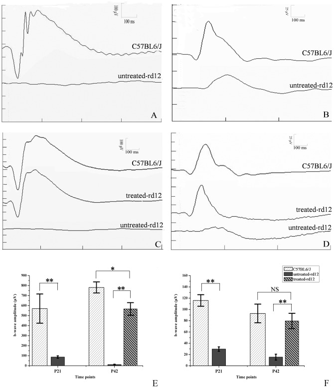
Figure 1. Scotopic and photopic ERG testing of untreated, treated rd12 and normal C57BL/6J eyes.
Panels A and B represent scotopic (panel A) and photopic (panel B) ERGs recorded on P21 from C57BL/6 (upper trace) or untreated rd12 mice (lower trace). Panels C and D represent scotopic ERG (panel C) and photopic ERG (panel D) recorded on P42 from C57BL/6 (upper trace), treated rd12 mice (middle trace) or untreated rd12 mice (lower trace). Panels E and F indicate statistical comparison between b-wave amplitudes of different groups at both time points under scotopic and photopic conditions, respectively. N = 6/group, and error bars depict the standard error of the mean. Scotopic ERGs (A and C) were obtained using white stimulus of −5 cd·s/m2, while photopic ERGs (B and D) were recorded using white stimulus of 1.96 cd·s/m2 of rd12 and C57BL/6J eyes. Scotopic ERG, b-wave amplitude of the untreated rd12 eye was 483.7±59.9 µV (p<0.001), lower than that of the normal control C57BL/6J eyes at P21, b-wave amplitude of the treated rd12 eye became 555.1±28.4 µV (p<0.001) on P42, higher than that of the untreated rd12 eye. In photopic ERG, b-wave amplitude gap was 86.2±4.54 µV (p<0.001) between the untreated rd12 and B6 eyes on P21, and was 64.2±6.5 µV (p<0.001) between the treated and untreated rd12 eyes on P42. NS: no significance. *: p<0.05; **: p<0.001.

