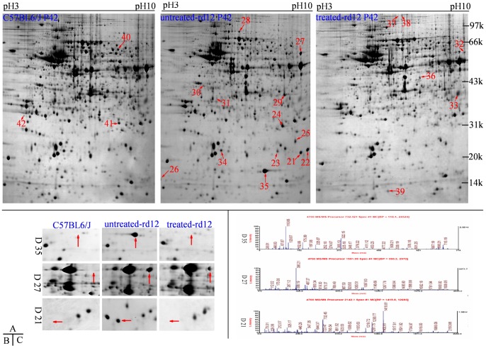
Figure 3. Differential retinal proteins at age P42.
Upper panels (A) represent 2-DE gels of wide-type C57BL/6J (left panel), untreated rd12 (middle panel) and treated rd12 eyes (right panel) at age P42. Data are a composite of 3 independent experiments, and n = 6/group. Differential proteins are marked with red numbers (from 21 to 42). Magnified details of differential proteins No. 21, 27 and 35 among the 3 eye groups at age P42 are presented in B. Graphical representation of mass spectrometry peaks from differential proteins No. 21, 27 and 35 are shown in C (the x- and y-axis represent mass-to-charge ratio (m/z) and relative intensity, respectively; the mass numbers of monoisotopic peaks [M+H]+ for peptides are marked above individual peaks).

