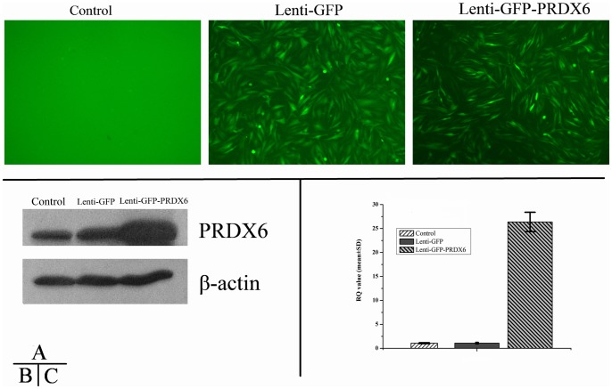
Figure 6. Immunofluorescence and Western blot of Lenti- PRDX6-GFP vector in MIO-M1 cells.
Transfection of Lenti-PRDX6-GFP was evaluated by immunofluorescence (A), western blot (B) and RT-PCR (C). Data are a composite of 6 independent experiments for A, and 3 for B and C. Panels in A represent confocal micrographs and demonstrate GFP immunofluorescence in transfected groups indicating successful transfection; Western blot results in B indicate that PRDX6 group expressed much more PRDX6 than either the GFP or the blank group at protein level; Bar graphs in C indicate the results from the RT-PCR analysis, demonstrating an over expression of PRDX6 6.3 times compared to the other two groups at mRNA level (p<0.01). Error bars depict the standard error of the mean. Calibration bar = 50 μm.

