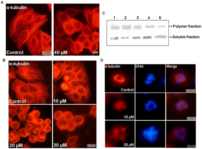
Figure 4. BCFMT depolymerized microtubules of MCF-7 cells.
(A) Cells were treated without and with 40 µM BCFMT for 3 h and microtubules were stained using antibody against α-tubulin (red). (B) BCFMT perturbed interphase microtubule organization of MCF-7 cells. MCF-7 cells were incubated in the absence and presence of different concentrations of BCFMT for 48 h. Cells were fixed and stained using antibody against α-tubulin (red). (C) BCFMT decreased the ratio of polymeric/soluble tubulin in MCF-7 cells determined by western blot. MCF-7 cells were treated without (lane 1) or with 20 µM (lane 4) and 40 µM (lane 5) of BCFMT for 36 h. 20 nM taxol (lane 2) and 200 nM nocodazole (lane 3) were also used under similar experimental conditions. Polymeric and soluble tubulin fractions were isolated and equal amounts of proteins were loaded on SDS-PAGE. Immunoblotting was done with α-tubulin antibody. Experiment was performed independently three times. Shown is the representative blot. (D) BCFMT depolymerized spindle microtubules in MCF-7 cells. DNA stained in blue. Scale bar is 10 µm.

